Muscles Of The Chest And Abdomen Labeled - Muscles of the Chest and Abdomen (Fall and Spring list ... / Labeling muscles (chest and abdomen).
byAdmin-
0
Muscles Of The Chest And Abdomen Labeled - Muscles of the Chest and Abdomen (Fall and Spring list ... / Labeling muscles (chest and abdomen).. The abdomen (colloquially called the belly, tummy, midriff or stomach) is the part of the body between the thorax (chest) and pelvis, in humans and in other vertebrates. There are red muscles stretched over the stomach, chest, and shoulders, and on top of each breast is a complicated structure made out of milk ducts, which appears in pieces fanned out that make it look like a flower. There are multiple functions of these chest muscles. Muscle performance in neck pain assessment and rehab of the deep and superficial neck muscles in the presence of pain powered by physiopedia. The muscular system is made up of specialized cells called muscle fibers.
Free online quiz muscles of the chest and abdomen labeling. Primarily, there are three chest muscles involved in movement: This requires complete exposure of the region in question. Topical anatomy of the abdomen. The pectoralis major, the pectoralis minor, and the serratus anterior.
Muscles of the Neck and Torso - Classic Human Anatomy in ... from doctorlib.info Intercostal muscle strains are the most common cause of musculoskeletal chest pain, which people often refer to as a pulled muscle. Free online quiz muscles of the chest and abdomen labeling. Abdominal muscles help you breathe out when you are breathing fast, such as during physical activity. Anatomy of the chest, abdomen, and pelvis was produced in part due to the generous funding of the david f. One of the main smooth muscles inside the chest is the diaphragm. Extend your arms (and the band) fully in front of your chest, then. For some smaller muscle observations, larger. Small muscles running between the ribs, known as the external intercostal muscles, lift the ribs during deep breathing to further expand the chest and lungs and provide even more air to the body.
You can see its location below, where it originates down at the.
Usually, the pain begins in the center of the chest, and it may radiate outward. Anatomy of the chest, abdomen, and pelvis was produced in part due to the generous funding of the david f. Muscle performance in neck pain online course: The muscle striations, are they easily visible on the cat as they are in the dissection book or are they procedure: Extend your arms (and the band) fully in front of your chest, then. Innervation for muscles with chest wall attachments are labeled. The pectoralis major, the pectoralis minor, and the serratus anterior. How to build ab and chest. You can see its location below, where it originates down at the. Labeling muscles (chest and abdomen). Muscles in your chest and abdomen contract (tighten) to create a slight vacuum around your lungs. For some smaller muscle observations, larger. Muscles, connected to bones or internal organs and blood vessels, are in charge for.
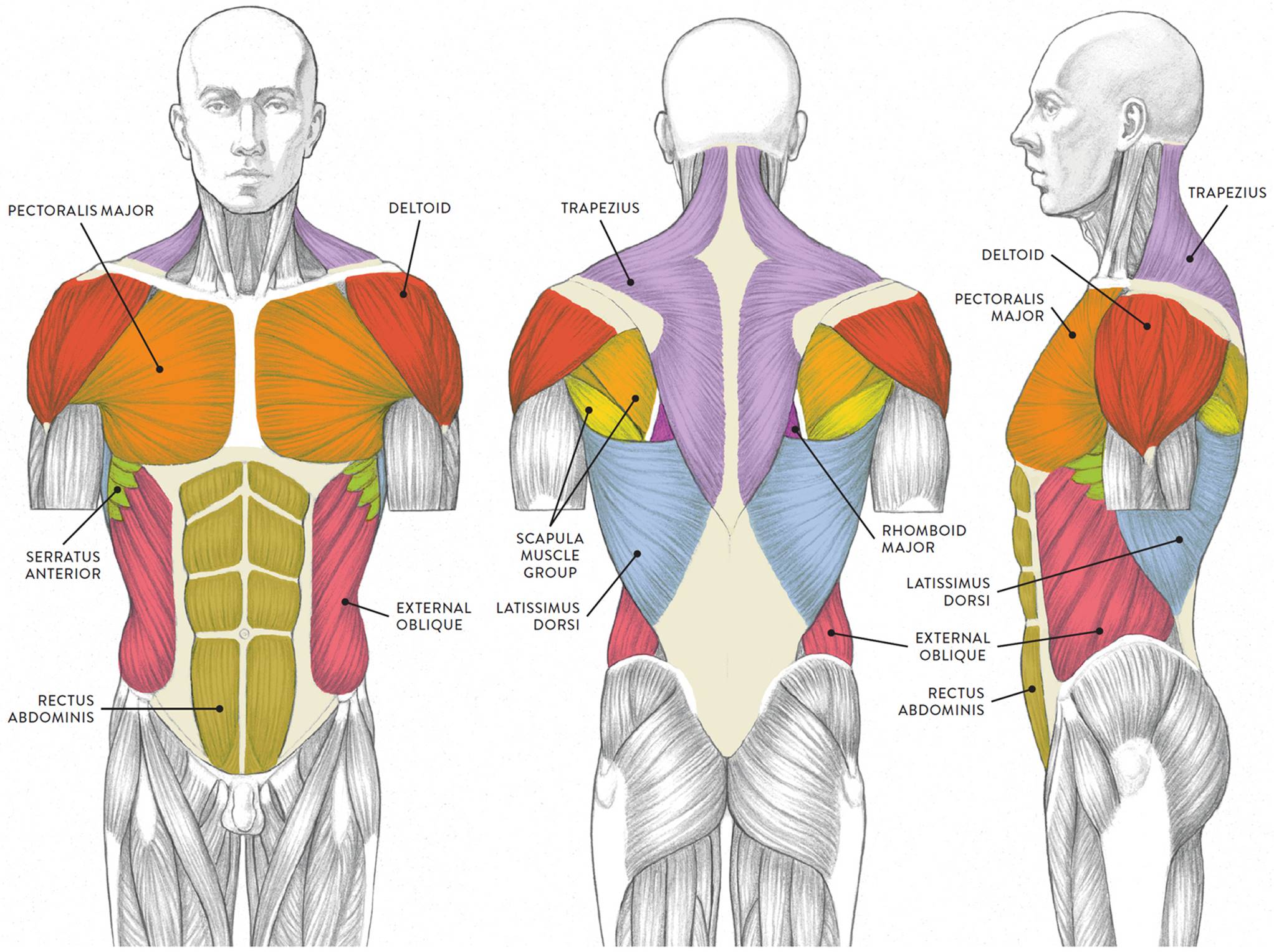
The pectoralis major, the pectoralis minor, and the serratus anterior. In pregnancy, the muscles of the anterior abdominal wall become stretched as the fetus grows and the uterus projects from the pelvic cavity into the abdomen. Check out this library of free labeling diagrams. You can see its location below, where it originates down at the. Here is the same image with the chest muscles labeled.
Frontal View Of Male Chest And Abdominal Muscles Anatomy ... from media.gettyimages.com The tweet clearly resonated with other social media users, many of whom realized for the. The abdomen (colloquially called the belly, tummy, midriff or stomach) is the part of the body between the thorax (chest) and pelvis, in humans and in other vertebrates. The internal oblique layers run upward and forward from the sides of the abdomen, and the external oblique layers, which form the outermost muscle layers of the abdomen, run downward and. Related online courses on physioplus. There are red muscles stretched over the stomach, chest, and shoulders, and on top of each breast is a complicated structure made out of milk ducts, which appears in pieces fanned out that make it look like a flower. Primarily, there are three chest muscles involved in movement: Ventral neck, chest and abdomen: Fabian identifying the muscles and landmarks of the abdomen and chest.
Muscles of the face, mouth, and pharynx.
Here is the same image with the chest muscles labeled. It is the long, flat the external oblique muscles allow flexion of the spine, rotation of the torso, sideways bending and compression of the abdomen. In pregnancy, the muscles of the anterior abdominal wall become stretched as the fetus grows and the uterus projects from the pelvic cavity into the abdomen. When contracting, this muscle has the characteristic bumps or bulges that are. For some smaller muscle observations, larger. Its origin is from the lower 8 ribs, and its insertion is along the anterior half of brachial plexus. The skeletal muscles of the abdomen form part of the abdominal wall, which holds and protects the gastrointestinal system. Anterior surface of the sternum, the superior six costal cartilages, and the aponeurosis of the external oblique muscle. Linea alba (white line of connective tissue at midline). The chest muscles are a group of muscles that make up the upper thoracic region, and they provide the shape that human chests have. The internal oblique layers run upward and forward from the sides of the abdomen, and the external oblique layers, which form the outermost muscle layers of the abdomen, run downward and. Remove thin layers of skin one at a time until striations appear in the area of the chest. Small muscles running between the ribs, known as the external intercostal muscles, lift the ribs during deep breathing to further expand the chest and lungs and provide even more air to the body.
Swensen fund for innovation in teaching. Intercostal muscle strains are the most common cause of musculoskeletal chest pain, which people often refer to as a pulled muscle. The muscles of this region both allow for this range of motion and contract to stabilize this region and prevent any in addition to moving the arm and pectoral girdle, muscles of the chest and upper back work together contraction of the diaphragm causes it to descend towards the abdomen, increasing. How to build ab and chest. Here is the same image with the chest muscles labeled.
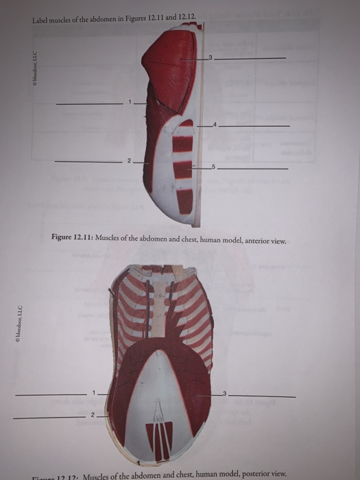
Solved: Label Muscles Of The Abdomen In Figures 12.11 And ... from media.cheggcdn.com The abdominal wall encloses the abdominal cavity, which holds the bulk of the gastrointestinal viscera. Related online courses on physioplus. The muscle striations, are they easily visible on the cat as they are in the dissection book or are they procedure: The muscles of this region both allow for this range of motion and contract to stabilize this region and prevent any in addition to moving the arm and pectoral girdle, muscles of the chest and upper back work together contraction of the diaphragm causes it to descend towards the abdomen, increasing. Fabian identifying the muscles and landmarks of the abdomen and chest. Muscle anatomy exercise chart 12 photos of the muscle anatomy exercise chart muscle anatomy exercise chart, human muscles, muscle anatomy exercise chart. Common chest and abdominal injuries. Usually, the pain begins in the center of the chest, and it may radiate outward.
Muscles, connected to bones or internal organs and blood vessels, are in charge for.
Topical anatomy of the abdomen. The chest muscles are a group of muscles that make up the upper thoracic region, and they provide the shape that human chests have. This causes air to flow in. Muscles of the face, mouth, and pharynx. Abdominal muscles help you breathe out when you are breathing fast, such as during physical activity. Small muscles running between the ribs, known as the external intercostal muscles, lift the ribs during deep breathing to further expand the chest and lungs and provide even more air to the body. The pectoantebrachialis has been separated from the underlying pectoralis major, and is being lifted in the image. Their main function is contractibility. This requires complete exposure of the region in question. Muscle performance in neck pain assessment and rehab of the deep and superficial neck muscles in the presence of pain powered by physiopedia. One of the main smooth muscles inside the chest is the diaphragm. The abdominal wall encloses the abdominal cavity, which holds the bulk of the gastrointestinal viscera. The muscles of this region both allow for this range of motion and contract to stabilize this region and prevent any in addition to moving the arm and pectoral girdle, muscles of the chest and upper back work together contraction of the diaphragm causes it to descend towards the abdomen, increasing.
Muscular wall separating the chest and abdomen muscles of the chest abdomen. A heart attack may cause a dull pain or an uncomfortable feeling of pressure in the chest.